Diseases and the Immune System in Animals
The immune system evolved over millions of years and our immune cells have learned to successfully fight diseases with incredibly fascinating strategies. Immunizations, for example, protect like a magic shield, making the body unassailable. Killer cells defend us like a secret army. We currently know nowhere near everything about this highly complex defense system and are learning more each day.
Here we describe some diseases and conditions that have been successfully treated in innovative immunological scientific studies. We know that many more diseases exist and will regularly be reporting new findings here. But please don’t forget that these new findings on the immune system have a direct and tremendous relevance not just for animals but also for humans.
Equine Sarcoids
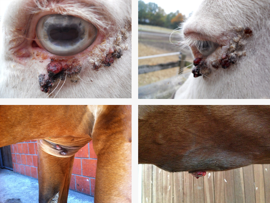
Equine sarcoids are the most commonly occurring skin tumours in horses. They are virus-induced pathological skin lesions that occur only externally and do not tend to metastasize to internal organs. After surgery, however, the tumour often recurs and large-area excision will mutilate the animal, e.g. loss of an eye.
Sarcoids are most common on areas of the body with little hair and thin skin such as the inside of the thighs, the face, and around the eyes and ears. Over 40% of all horses carry this virus. There is no breed, gender or color predilection. Equine sarcoid is more than a cosmetic problem. If the location is unfavorable, it spreads, or it is improperly treated, the horse may become unusable or even require euthanasia.
Causes
The cause of equine sarcoid is the bovine papilloma virus (BPV Type 1 and 2). The tiniest wounds in the skin can be the site of entry of the virus. Usually the skin tumours are found in animals between the ages of three and seven. The DNA of the virus can be identified in nearly 100% of all tumours. Parts of the virus genome can also be identified in the blood of horses. Evidently, an inadequate immune defense is responsible for onset of the disease. Also genetic predispositions or a combination of these factors may be involved.
Tumour Types
There are six different classifications according to Pascoe and Knottenbelt:
- Type 1: Occult sarcoid: The affected skin is usually hairless and slightly raised and sometimes there is slightly nodular and hyperkeratotic growth.
- Type 2: Verrucose sarcoid: It appears as a wart-like growth with hyperkeratosis.
- Type 3: Nodular sarcoid: It presents as subcutaneous nodules of varying size and can be pedunculated. The skin above it is usually hairless but intact.
- Type 4: Fibroblastic sarcoid: It has a cauliflower-like appearance with ulcerated surface and serous exudates. It may also have a pedicle (stalk).
- Type 5: Mixed sarcoid: It looks like a mix of the verrucose, nodular and fibroblastic sarcoids.
- Type 6: Malevolent sarcoid: It often appears after multiple traumas and can infiltrate the lymph system. Then nodules can be seen along the lymph channels.
The mild types named above first can transform to the serious types after removal, but also spontaneously.
Treatment
Many treatment options are available for therapy of equine sarcoid, but they are usually too ineffective or costly. Surgical excision poses the risk that not all parts of the tumour are removed, which would result in recurrence. Also used is chemotherapy or immunization with Bacillus Calmette-Guérin, a live vaccine against tuberculosis. Often different forms of therapy are combined. With a cure rate of over 80% to 90%, radiotherapy – either as brachytherapy or teletherapy – offers the best chances for complete recovery. Brachytherapy has been available in German since 2009, but is used very rarely because of the high costs. Besides harming the animal, the economic damage caused by the papilloma virus is considerable. The use, sale and breeding of the horse are threatened and there is a great need for effective, safe, easy to use and affordable therapies.
Outlook
4 Animals AlsterScience GmbH works to directly influence the defense against the pathogen and is currently developing therapies that improve the immune function of animals and support their immunological defense.
Strengthening specifically of the immune system is an innovative approach. The body’s own proteins ensure that disease-causing pathogens can be repelled without use of antibiotics or virostatics. Especially in veterinary medicine, where infections can spread quickly in groups, there is a tremendous need to improve resistance to pathogens in order to avoid follow-up therapies (e.g., antibiotics, operations, radiation treatments). Infections, tumours, stress and demands for high performance are regularly associated with disturbances in the function of the immune system; viruses can multiply successfully and impair immune system function by infection. This results in a vicious circle with worsening of the course of the disease and long-term complications.
4 Animals AlsterScience GmbH is currently working closely with recognized experts to accomplish in effective steps the development of highly effective, well-tolerated products for pets and food-producing animals that strengthen the immune system.
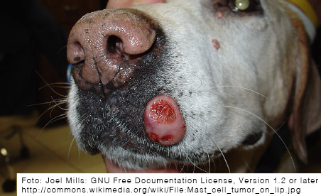
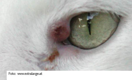
in dogs and cats